DynaMo Seminar: Dorus Gadella
DynaMo Center of Excellence is pleased to invite you to our next DynaMo Seminar with Professor Theodurus Gadella from the University of Amsterdam:
Enhanced fluorescent proteins for FRET and for studying signaling across the membrane
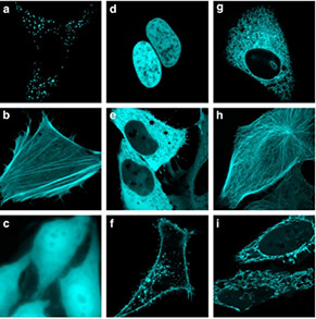
 Since the cloning of the green fluorescent protein from Aequoria victoria, numerous fluorescence microscopy applications have been described in the literature employing new GFP variants. Here we report a screening method that, in addition to fluorescence intensity, quantifies the excited state lifetime of a fluorescent protein, providing a direct measure for the quantum yield of the fluorescent protein. The novel approach was used to screen a library of cyan fluorescent protein (CFP) variants yielding a very bright cyan fluorescent protein variant, which we dubbed mTurquoise. mTurquoise has a marked increased quantum yield of 0.84, and a seriously increased lifetime of 3.7 ns. After crystallization of mTurquoise one suboptimal residue Ile146 was identified that causes partial collisional quenching. Substitution with a phenylalanine (I146F) enhanced the fluorescence quantum yield to 0.93 (a record for monomeric fluorescent proteins). This mutant dubbed mTurquoise2 is the preferred donor to YFP variants (like mVenus or mCitrine) with an R0 of 5.83 nm.
We apply the different probes for the study of GPCR-triggered signaling across the membrane in single mammalian cells. Using mTurquoise, a new Gq FRET sensor was developed reporting on activation of the G protein by endogenous expressed receptors. Also downstream signaling events like multiparametric imaging of lipid-derived second messengers, PtdInsP2-dependent PLC relocalization, and RhoGEF activation using a variety of FRET-, ratio imaging- and TIRF-microscopic applications will be presented.
Dr. Theodorus Gadella is co-founder/director of the van Leeuwenhoek Centre for Advanced Microscopy (LCAM) and full professor in Molecular Cytology chairing a group of 25-30 scientists at the University of Amsterdam. The LCAM is a joint advanced microscopy centre between the Science Faculty of the University of Amsterdam, the Academic Medical Centre (AMC) and the Netherlands Cancer Institute (NKI-AVL) in Amsterdam. Recently LCAM was expanded to harbour the Nikon Centre of Excellence on Super Resolution Microscopy Development. Dr Gadella personally supervises a research team on spatiotemporal cell signaling. His team wants to understand how cells can achieve and maintain a local signal in order to drive morphogenesis, to define new cytoskeletal anchorage or vesicle-docking sites. The focus is on signal flow across and in the plane of the membrane of living mammalian cells. To this end genetic encoded fluorescent biosensors are developed and employed, and the in situ molecular interactions between signaling molecules including phospholipid-second messengers, receptors, G-proteins and downstream targets are analyzed. For these biosensors new superfluorescent proteins have been developed such as SYFP2 (the brightest GFP variant on the planet) and mTurquoise2 (the brightest cyan fluorescent protein known to date). By multiparameter imaging approaches several signaling events are visualized and quantified simultaneously in individual living cells with sub-second temporal and submicron spatial resolution. The in situ cellular imaging research heavily depends on advanced bioimaging. To this end advanced automated microscopy approaches such as fluorescence resonance energy transfer (FRET) microscopy (including bleaching-, spectral-, ratio- and fluorescence lifetime-imaging approaches), fast live cell microscopy (including spinning disk, line-scanning and controlled light exposure confocal microscopy, and total internal reflection microscopy (TIRF)), and photochemical microscopy approaches such as photoactivation, uncaging, fluorescence recovery after photobleaching (FRAP), fluorescence loss in photobleaching microscopy (FLIP), fluorescence correlation spectroscopy (FCS), cross-correlation (FCCS), lifetime-correlation (FCLS) have been implemented developed and applied to quantifying cell signalling phenomena. Recently, the palette of advanced imaging instrumentation is expanded with superresolution localization microscopy approaches such as SIM, PALM and STORM. In 2007 prof Gadella was elected president of the Netherlands Society for Microscopy. Recently he has become national coordinator of the NL-BioImaging Advanced Microscopy consortium of 18 collaborating advanced microscopy centres in the Netherlands that connects to the new large EU infrastructure (ESFRI) roadmap Euro-BioImaging that entered the preparatory phase in 2010.
----
Goedhart J, von Stetten D, Noirclerc-Savoye M, Lelimousin M, Joosen L, Hink MA, van Weeren L, Gadella TWJ, Royant A. (2012) Structure-guided evolution of cyan fluorescent proteins towards a quantum yield of 93%. Nat Commun. 3:751.
Goedhart J, van Weeren L, Adjobo-Hermans MJ, Elzenaar I, Hink MA, Gadella TWJ (2011) Quantitative co-expression of proteins at the single cell level--application to a multimeric FRET sensor. PLoS One. 6(11):e27321.
Goedhart J, Gadella TWJ (2011) Bypassing GPCRs with chemical dimerizers. Chem Biol. 18(9): 1067-1068.
Adjobo-Hermans MJ, Goedhart J, van Weeren L, Nijmeijer S, Manders EM, Offermanns S, Gadella TWJ (2011) Real-time visualization of heterotrimeric G protein Gq activation in living cells. BMC Biol. 9:32.
Klarenbeek JB, Goedhart J, Hink MA, Gadella TWJ, Jalink K. (2011) A mTurquoise-based cAMP sensor for both FLIM and ratiometric read-out has improved dynamic range. PLoS One. 6(4):e19170.
Goedhart J, van Weeren L, Hink MA, Vischer NO, Jalink K, Gadella TWJ (2010) Bright cyan fluorescent protein variants identified by fluorescence lifetime screening. Nat Methods. 7:137-139.
Speaker
 |
Prof. dr. T.W.J. Gadella
Section of Molecular Cytology, University of Amsterdam
